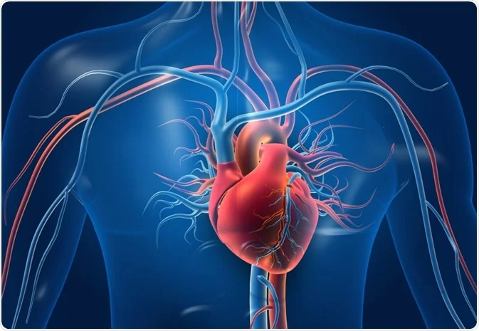
The human heart is a muscular organ that pumps blood throughout the body. It is roughly the size of a fist and is located in the chest, slightly to the left of the midline. The heart is divided into four chambers and works as a dual pump, sending oxygen-rich blood to the body and oxygen-poor blood to the lungs.
Anatomy of the Human Heart
1. Chambers of the Heart
The heart has four chambers:
- Right Atrium – Receives oxygen-poor blood from the body through the superior and inferior vena cava.
- Right Ventricle – Pumps oxygen-poor blood to the lungs via the pulmonary artery.
- Left Atrium – Receives oxygen-rich blood from the lungs through the pulmonary veins.
- Left Ventricle – Pumps oxygen-rich blood to the body through the aorta.
2. Valves of the Heart
To ensure unidirectional blood flow, the heart has four valves:
- Tricuspid Valve – Between the right atrium and right ventricle.
- Pulmonary Valve – Between the right ventricle and pulmonary artery.
- Mitral (Bicuspid) Valve – Between the left atrium and left ventricle.
- Aortic Valve – Between the left ventricle and aorta.
3. Major Blood Vessels
- Superior and Inferior Vena Cava – Carry oxygen-poor blood from the body to the right atrium.
- Pulmonary Arteries – Carry oxygen-poor blood from the right ventricle to the lungs.
- Pulmonary Veins – Carry oxygen-rich blood from the lungs to the left atrium.
- Aorta – The largest artery, carrying oxygen-rich blood from the left ventricle to the body.
4. Layers of the Heart Wall
- Endocardium – The inner lining of the heart.
- Myocardium – The thick muscular layer responsible for contractions.
- Epicardium – The outermost protective layer.
5. Blood Circulation in the Heart
- Pulmonary Circulation – Blood moves from the right side of the heart to the lungs for oxygenation and back to the left side.
- Systemic Circulation – Oxygen-rich blood is pumped from the left side to the entire body and returns oxygen-poor blood to the right side.
6. Electrical Conduction System
The heart’s rhythmic contractions are controlled by electrical signals:
- Sinoatrial (SA) Node – The natural pacemaker of the heart, located in the right atrium.
- Atrioventricular (AV) Node – Delays the impulse before sending it to the ventricles.
- Bundle of His & Purkinje Fibers – Help distribute the electrical impulse to ensure coordinated contraction.
