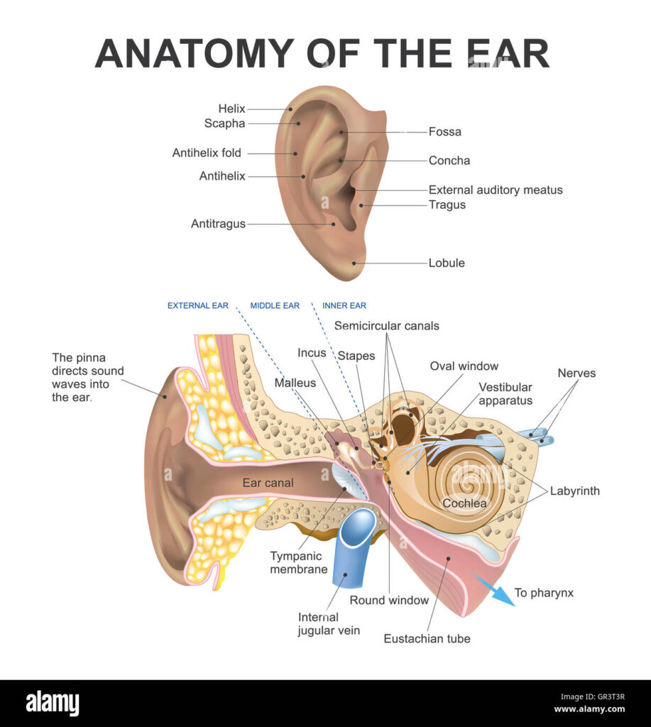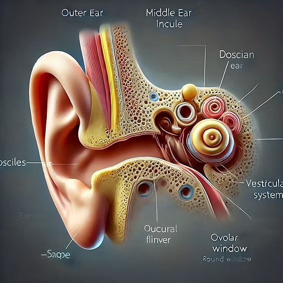
The anatomy of the ear is divided into three main parts: the outer ear, middle ear, and inner ear. Each part plays a critical role in hearing and balance.
1. Outer Ear (External Ear)
- Auricle (Pinna): The visible part of the ear that collects sound waves and directs them into the ear canal. It also helps in determining the direction from which sound is coming.
- External Auditory Canal (Ear Canal): The passage that carries sound waves from the auricle to the eardrum. It also protects the inner parts of the ear from debris and infection.
2. Middle Ear
- Tympanic Membrane (Eardrum): A thin membrane that vibrates when sound waves hit it. These vibrations are transferred to the bones in the middle ear.
- Ossicles: Three small bones that transmit the vibrations from the eardrum to the inner ear. They are:
- Malleus (Hammer): Attached to the eardrum and connects to the incus.
- Incus (Anvil): Connects the malleus to the stapes.
- Stapes (Stirrup): Connects to the oval window, transmitting vibrations to the inner ear.
- Eustachian Tube: A tube that connects the middle ear to the throat, equalizing pressure in the middle ear, which is essential for proper hearing.
3. Inner Ear
- Cochlea: A spiral-shaped organ responsible for converting sound vibrations into nerve signals. It is filled with fluid and contains hair cells that respond to sound vibrations, which are then sent to the brain via the auditory nerve.
- Vestibular System (Semicircular Canals): A set of three fluid-filled tubes that help with balance and spatial orientation. They detect the rotation of the head.
- Oval Window: A membrane-covered opening that connects the middle ear to the cochlea, where the vibrations are transmitted from the stapes.
- Round Window: Another membrane-covered opening that allows for the release of pressure within the cochlea.
These structures work together to allow us to hear and maintain balance. The auditory signals are processed by the brain to create the perception of sound, while the vestibular system helps with equilibrium.

